The Shroud of Turin · Chapter 7 of 21
The digital autopsy: reconstructing the body from the image
If the image on the Shroud encodes distance, then in principle the image can be turned into a depth map, and the depth map can be fitted to a model of a human body. Such a fitted model is sometimes called a digital autopsy: a way of reading the cloth as a record of the body that lay in it. The idea is attractive, and it has produced some of the most striking images of the cloth. It is also easy to overstate. This chapter sets out what digital analysis can show, how the methods work and where each stage can change the result, what the facial reconstructions do and do not establish, and what a study would need before its results could be treated as evidence about the man in the cloth.
The foundation: what the earlier work established
Digital reconstruction rests on a chain of earlier findings, and each link in the chain has its own history. The first is Pia’s 1898 discovery that the body image is a photographic negative, so that a positive view of the image shows a lifelike figure.1 The second is the 1978 finding that the image coloration is confined to the outermost fibrils of the linen, with no detectable pigment, dye, or binder.2 The third is the relationship between image density and modelled cloth-to-body distance, reported first by the VP-8 analysis of 1976 and then quantified by Jackson, Jumper, and Ercoline in 1984.3 The fourth is the independent confirmation of the relief by Giovanni Tamburelli, who processed the 1931 photographs digitally in 1981.4 Later work reported that the image’s information is concentrated in a single intensity variable, as a principal component analysis of the 2002 photograph found in 2025.5
Taken together, these results show that the image’s tonal structure is systematic and that it has been reproduced by several analysts. They do not show that the structure is a body, nor that the body can be recovered in any detail. Digital reconstruction is an attempt to go from the structure to the body, and the gap between the two is where most of the disagreement lies.
The limits, stated at the outset
Before describing what a reconstruction can show, it is necessary to say what it cannot. The statement is not a disclaimer. It is the thing that makes the positive claims credible. A reconstruction cannot date the cloth. The radiocarbon question is a materials-science question, separate from the morphology of a face or a body, and the morphology does not settle it. A reconstruction cannot explain how the image was formed, because it takes the intensity pattern as given. And a reconstruction cannot confirm the identity of the man. A coherent human form, fitted to the data, may be anatomically consistent and internally coherent. Whether it represents the historical man is a separate question, answered by historical and textual evidence operating by different methods.6
What a reconstruction can offer is narrower and more checkable. It can show whether the encoded data is structurally coherent and anatomically plausible. It can show whether the body it models is internally consistent across its measurable dimensions. And it can show whether the wounds it places are spatially consistent with a body in a particular position, rather than scattered marks. Those are useful results, and they are the results a careful study should aim to produce.
The pipeline
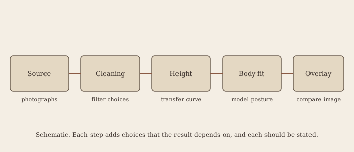
A digital reconstruction moves through several stages. Each adds assumptions, and each can change the result. The table below sets out the stages, what each does, and what it can alter.
| Stage | What is done | What it can change |
|---|---|---|
| 1. Source images | Choosing the photograph: the 1898 Pia plates, the 1931 Enrie prints, the 1978 STURP photographs, or the 2002 Durante photograph | Each source has different lighting, resolution, exposure, and damage history |
| 2. Cleaning | Reducing the herringbone weave, masking the 1532 fire scorches and water stains, and removing repairs and damage | Filtering can remove real structure or create false structure |
| 3. Height conversion | Mapping image intensity to a value interpreted as height or distance | The mapping is a choice, and the output depends on it |
| 4. Body fitting | Fitting a body model to the relief, usually with draping and posture assumptions | Posture, landmark choice, and the model’s proportions |
| 5. Detail addition | Adding skin, hair, wounds, or a face | Anything added at this stage is not in the image data |
| 6. Checking | Overlaying the model on the image and comparing | A visual match can be produced by an adjusted model, so an overlay does not prove the model |
Stages one to four are where measurement is possible, and where the work can be checked against the photographs. Stage five is where the output stops being a measurement and becomes an interpretation. Stage six is a consistency check, not an independent test. A reader who wants to weigh a reconstruction should ask, for each stage, what was measured, what was assumed, and what was added.
Source images
The choice of source image matters more than it might seem. The 1898 plates are the earliest and the most direct record of the negative effect, but they were made with equipment that is poor by modern standards, and their surviving copies vary in contrast and tone. The 1931 Enrie prints are of higher technical quality, and they have been the basis of most of the mid-twentieth-century analysis. The 1978 STURP photographs include colour, ultraviolet, and infrared records, which are valuable for the blood and the image separately. The 2002 Durante photograph is the highest-resolution full-image photograph in common use, and it has been the basis of the recent statistical work.
A reconstruction should state which source it used, and it should say whether the same results appear with other sources. A result that depends on one photograph, with one contrast curve and one processing chain, is less robust than one that appears in several.
Cleaning and its dangers
The cleaning stage is designed to isolate the body image from the damage and the weave. The herringbone pattern of the cloth is a periodic structure, and it can be reduced by filtering. The scorches and holes of the 1532 fire can be masked, and the water stains can be reduced. Each of these operations is reasonable, and each can change the result. A filter that removes the weave can also remove real structure if the structure has a similar spatial scale. A mask over the scorches can remove a region that carries information. The processing has to be recorded, and its effect on the output should be tested by varying the filter settings.
A feature that disappears when the filter is changed is not a robust anatomical signal. A feature that survives a range of reasonable filter settings has a better claim to be a property of the image. That is a basic test, and it is often omitted from published reconstructions.
Height conversion
The height conversion maps image intensity to a value interpreted as height or distance. This is the most consequential choice in the pipeline, because it defines the relation between brightness and geometry. The VP-8 used a fixed transfer function, and that function determined the output. Later work has used linear mappings, nonlinear mappings, and mappings fitted to the physical distance model. Each mapping gives a different surface, and the surface can change in its details, its proportions, and its apparent relief.
The conversion also depends on the physical model of distance. If the image intensity falls off with distance in a particular way, the mapping should reflect that. Jackson and his colleagues fitted a global function across the frontal image, and they acknowledged that the function was an approximation.3 The approximation is a choice, and a reader should ask whether another function would produce a different result.
Body fitting and the question of stature
Once a relief has been produced, a body model can be fitted to it. The model may be a computer-generated human, an articulated manikin, or a parametric body. The fit requires assumptions about posture, about the position of the head, knees, and feet, about the draping of the cloth over the body, and about the proportions of the model. Each assumption can shift the result.
Stature is the most studied output of body fitting. The Jackson and Jumper model gave a height of about 178 to 180 centimetres.3 A later analysis by Fanti, Basso, and Bianchini, which used a computerised manikin with cloth-wrapping corrections and posture allowances, gave about 175 centimetres, with a margin of about two centimetres.7 The two figures are close, and they rest on different assumptions. The result supports a broadly consistent adult stature. It does not support a measurement precise to one or two centimetres.
The frontal and dorsal images differ in length. The frontal image measures about 1.95 metres and the dorsal about 2.02 metres. Those lengths cannot be read directly as stature, because the cloth is not a rigid shell and because the head, knees, and feet are positioned differently in the two images. The manikin analysis showed that the two images can be made compatible with one body under plausible assumptions. That is a demonstration of consistency, not a proof that both images show a single body.
Detail addition: where the data ends
The fifth stage adds detail, and it is the stage where the reconstruction most easily goes beyond the evidence. Skin texture, the shape of the lips, the colour of the hair, and the precise form of the face are not encoded in the intensity map in the way that the broad shape of the body is. They are supplied by the artist or by the software that generates them. Some reconstructions have used generative artificial intelligence tools trained on large collections of modern photographs. Such tools produce convincing detail, and that detail is a product of the training data and the prompt, not of the cloth.
The distinction should be kept clear. A relief, fitted to a body model, is a constrained inference from the image. A face generated to fill that relief is an interpretation. The two should be reported separately, and the interpretation should not be presented as data. A reconstruction that blurs the line between them has stopped being a measurement.
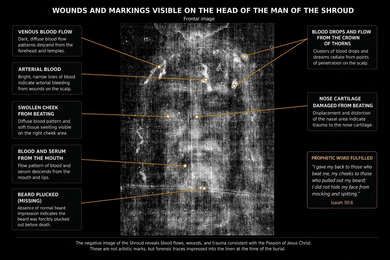
Wounds added to a model
Some reconstructions add wounds to the model at the places where the cloth shows marks, and then use the wounds as a check on the body’s proportions. The logic is that a blood path running down the model’s head should follow the head’s shape, and that a scourge mark should fall on a plausible part of the back. The logic is sound only if the wounds are taken from the cloth and placed without adjustment.
When wounds are painted or inpainted by an artist or a tool, the test becomes circular. The model is adjusted to fit the wounds, and the wounds are then cited as confirmation of the model. Wounds added by that route are interpretation. They cannot be counted as evidence on the same footing as the measured relief, and a careful reader will ask at each stage whether a feature was measured or placed.
Overlays and their limits
A common check places a reconstructed body over the image at partial transparency and asks whether the features line up. The nose should fall in the darkest region, the eye sockets in lighter regions, and the wounds should follow the gravity paths of the blood. Colour-channel overlays make small mismatches visible, and they are a useful way of finding errors in a model.
The overlay is a test of consistency, and it cannot separate data from invention. A model can be adjusted until its features fit the image, and an artist who draws a face can make it fit a dataset that guided the drawing. A good overlay does not show that the detail came from the image. It shows only that the model and the image are compatible at the points checked, and the number of points checked is usually small.
The face
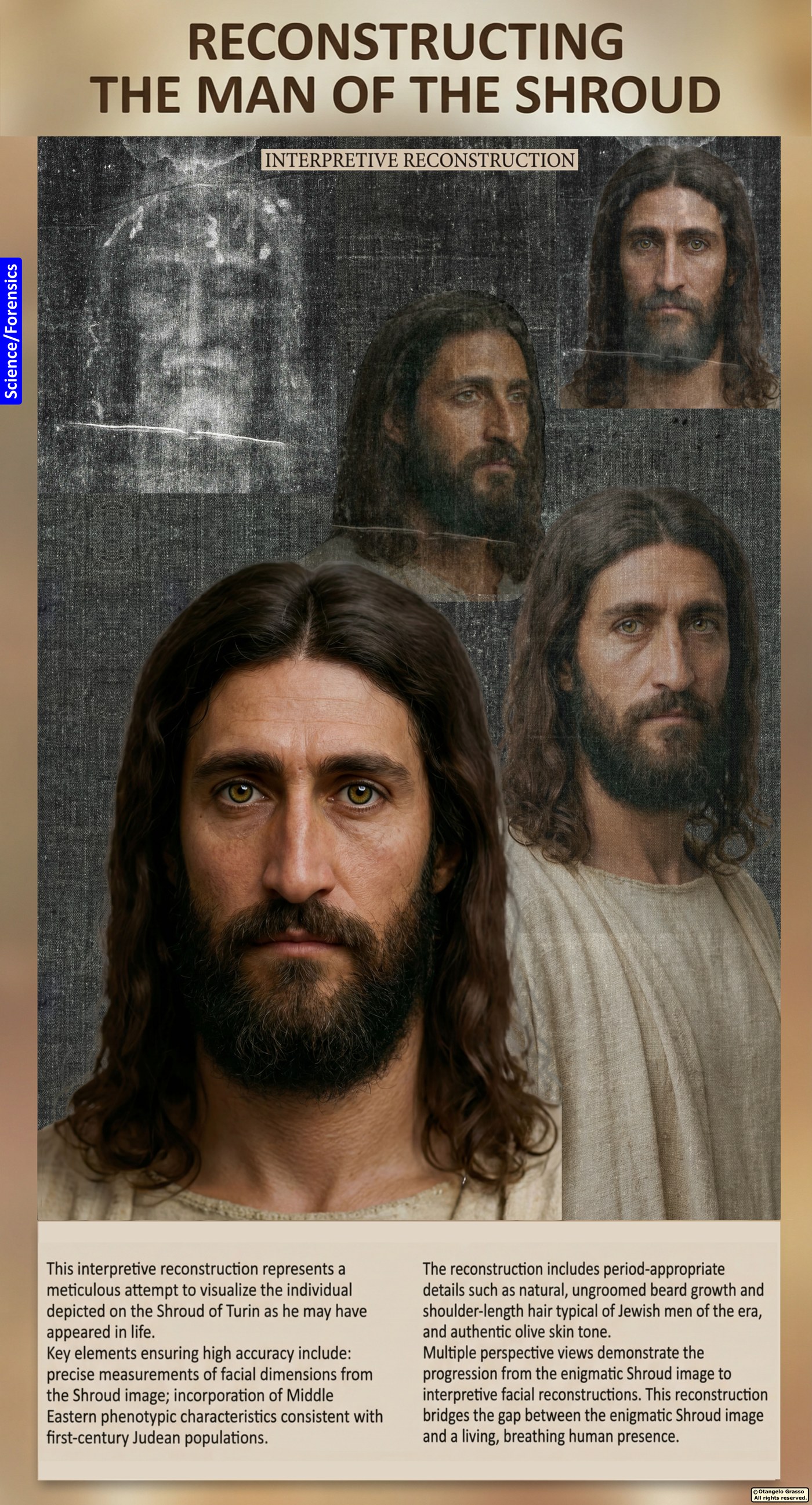
The most public outputs of digital reconstruction are faces. Several have been published, some as AI-generated portraits described as based on Shroud data. They attract attention because they show what a viewer expects a face to look like, and because the detail they contain is supplied largely by the generator.
There are two separate problems. The first is methodological. A depth-like map of the cloth can show the broad shape of the head, but the features that matter most to recognition, such as the exact shape of the nose and the form of the eyelids, depend on details that the image does not measure reliably. The second is pastoral. An image of Jesus generated by software and presented as the face of the Shroud risks putting an artefact in the place of the person. The image of the cloth is best read as physical evidence about a man who suffered and died, not as a portrait that the viewer is invited to complete.
The panel at the head of this chapter is an interpretive reconstruction. It should be read as an artist’s informed model, not as a measurement of the face.
An example of a documented reconstruction
Otangelo Grasso has published an AI-assisted reconstruction of the Shroud figure, which documents a workflow from image preparation through overlay checks, colour-channel comparison, and wound integration. It treats the cloth as the fixed reference that the model must satisfy.8 The work is self-published and has not been independently reviewed. Its own text acknowledges that generative tools can introduce anatomical conventions that are not in the cloth, and that the overlay cannot fully separate what the cloth encodes from what the tool supplies. It is a useful example of a method, and it should be read as an interpretation rather than as a measurement of the man.
What a credible study would need
A reconstruction that aims to be evidence needs more than a convincing image. The following conditions would make its results testable.
- Raw data first. The height conversion and the body model should be applied to minimally processed images, and the results should be reported with and without each filter and each transfer function.
- Full-image analysis. The frontal and dorsal images should both be analysed. The 1984 correlation used thirteen facial points, and it has not been replicated across the whole image with modern body models.3
- Independent replication. Another group should run the same pipeline on the same data, and report the outcome, including failures.
- Principal-component preprocessing. Reducing the image to its dominant intensity variable before correlation would make the analysis less sensitive to colour and channel choices.5
- Falsifiable predictions. A draped body should produce lateral distortion at the flanks, and a relief should not produce image in the hollows of the eyes. Each can be checked against the image.9
- No generated detail in the data. Any model built from the image should be kept separate from detail added by a generator, and the two should be reported separately.
- Stated sources. Each photograph used should be named, with its date, its processing, and its provenance.
Moraes, Casabianca, and the bas-relief question
The most important objection to the depth reading is that the image may have been made from a relief rather than a draped body. In 2025 Cicero Moraes used open-source body models and fabric simulation to compare a full body with a low-relief model. He reported that the low-relief model gave a contact pattern closer to the image, with less distortion.10 Casabianca, Marinelli, and Piana answered that the study modelled only the frontal image, treated contact as the only possible mechanism, and did not address the chemistry of the image. Moraes replied that his scope was limited to contact geometry.11
The geometric debate is open. The chemical question is separate. A contact model that fits the geometry still has to account for the superficiality of the colour, the absence of pigment, and the fact that the blood was on the cloth before the image formed. A digital reconstruction that does not address those points has answered a narrower question than the one it claims to address.
A short history of reconstruction
The idea that the image could yield a body is older than digital tools. Pia’s 1898 negative made the figure legible as a human form, and the early twentieth-century students of the cloth, such as the anatomist and historians who studied the Enrie prints, read the wounds and the figure as the record of a body. Their reading was visual and anatomical, not computational. It was limited by what the eye could resolve on a photographic print, and by the assumptions the readers brought to the picture.
The first instrumental reconstruction came with the VP-8 in 1976. The machine did not construct a body. It produced a height map of the face, and the map was then interpreted by the observers. The 1984 study introduced a quantitative model of draped linen and a fitted body, and it set the template for later work by making the relation between density and distance explicit.3 From that point the reconstruction became a question of model-building, and the models have grown more elaborate since.
The 1980s and 1990s brought digital image processing and more sophisticated fitting, and the 2000s brought high-resolution scanning and enhancement. Petrus Soons, working with enhanced versions of the 1931 photographs in the 2000s, produced depth data and holographic prints of the face, and his work showed the potential of modern enhancement.12 The 2010s and 2020s added fabric simulation, statistical preprocessing, and the generative tools that now make it easy to produce a convincing face. Each stage has added capability, and each has added assumptions. The history is a history of growing power and growing difficulty in separating what is measured from what is supplied.
What the reconstructions have agreed on
Across the decades, several conclusions recur in the reconstruction literature. The face in the image is broadly consistent with a head of an adult male, with a beard and longer hair. The shoulders, chest, and arms are consistent with the frontal and dorsal images. The stature, in the range of the published estimates, is broadly adult and not extreme. The wounds described in the forensic literature, such as the scalp punctures and the scourge marks, have been placed in positions that the body model accommodates. And the relief is coherent across different processing chains, although its fine detail varies.
These are useful points of agreement. They are also points that can be reached with modest assumptions, and they do not depend on any single contested result. The disagreements are about detail, about the position of the body, and about the relation between the relief and the physical body. Those are the points on which the reader should be most careful.
Where the reconstructions disagree
The reconstructions disagree on several points, and each disagreement is informative. They disagree on the precise stature, because they use different landmarks and posture assumptions. They disagree on the shape of the chest and the abdomen, because the cloth draping over the torso is poorly constrained. They disagree on the position of the head, because the head is tilted in the frontal image and the tilt is difficult to model. And they disagree on the degree of relief in the dorsal image, where the processing is more difficult and the image is less clear.
The disagreements suggest that the detailed anatomy of the figure is less secure than the broad form. A reader who sees a reconstruction with a precise nose, a precise jawline, or a precise head angle should ask whether those features are in the image data at the level of precision claimed, or whether they have been supplied by the model or the artist.
A checklist for reading a reconstruction
The following questions help when assessing any reconstruction of the Shroud figure. Which photograph was used, and was it processed? Which height conversion was applied, and was it tested with other conversions? Which body model was fitted, and how were its proportions chosen? Were the posture and draping assumptions stated and varied? Were the landmarks chosen before the result was known? Was the frontal image analysed alongside the dorsal, and were the two compatible? Were any features added to the model, and if so, were they separated from the measured data? Were the results replicated by an independent group? And does the reconstruction state its own limits clearly?
A reconstruction that answers these questions openly is more useful than one that shows an attractive image without the method. The checklist applies to the work of anyone who publishes a reconstruction, including those who favour the authenticity of the cloth and those who oppose it. The aim is to make the result testable, whatever its direction.
The ethics of the face
The face of the figure has a special status in the reconstruction literature. It is the most legible part of the image, and it is the part that viewers most want to see. It is also the part that carries the strongest religious associations, because the figure is widely identified with Jesus of Nazareth. A reconstruction of the face therefore raises questions that go beyond method.
The theological concern is that an image of the face, produced by a computer or an artist, may come to replace the person it is supposed to represent. The reconstruction becomes an object of devotion, and the viewer comes to believe that science has supplied what faith was meant to supply. The concern has been raised by writers on both sides of the authenticity question, and it applies to the scientific community as much as to the devotional one. A reader who holds the Gospel account can recognise the force of the concern without rejecting the study of the image.
The scientific concern is that a convincing face may be taken as evidence, and that the viewer may forget how much of the face was supplied by the generator. A face that looks like a photograph is persuasive, and persuasion is not the same as measurement. A responsible presentation of any reconstruction should say which parts are measured, which are inferred, and which are generated.
Cross-checks available to the forensic readings
The reconstruction is not the only means of checking the forensic readings. The wounds described in the forensic literature can be compared with clinical knowledge of crucifixion and scourging, with the Gospel account, and with the blood chemistry and flow patterns discussed in Chapter 3. A reconstruction that places a wound in a particular position should be consistent with those other sources. If it is not, the reconstruction is wrong, or the reading of the wound is.
The body proportions can also be checked against the archaeological record of the period, and against the skeletal evidence for stature. The archaeological estimates are imperfect, but they set a range within which a reconstruction should fall. A reconstruction that places the body far outside that range should be treated with caution, though a figure at the edge of the range is not thereby excluded.
What the reconstruction can contribute to the series
The reconstruction contributes a set of constraints, and a set of questions. It constrains the body to be of broadly adult stature, with proportions consistent with the frontal and dorsal images, and with wounds placed in positions that the forensic readings support. It raises questions about the depth of the relief, about the dorsal image, about the draping of the cloth, and about the detail of the face. Those questions shape the agenda of the next stage of work, which is set out below.
What a next stage of work would test
A next stage of work would test three things. The first is whether the relief survives a systematic variation of the processing, including the height conversion, the filter, and the photograph. The second is whether the frontal and dorsal images give a compatible body under a range of posture and draping assumptions, with the assumptions stated in advance. The third is whether the wounds, when placed without adjustment, fall in positions that the body model supports. Each test can be carried out with existing photographs and existing methods, and each would move the discussion from illustration toward measurement.
Such a stage of work would also need a clear statement of what would count as failure. A reconstruction that cannot be replicated, that depends on one processing choice, or that requires adjustments to fit the wounds, has failed the test, whatever its visual appeal. A fair study states its failure conditions before it begins, and it reports them honestly when they are met.
The lesson of the worked example is practical. A reader who wants to judge a reconstruction should not look first at its most attractive feature. The reader should look at the stages that produced it, identify the choices, and ask which features survive when the choices are varied. A reconstruction that holds its main features across a range of reasonable choices is a stronger piece of evidence than one whose features depend on a single chain of processing, however clear the final picture looks.
For that reason the series treats the reconstruction as a tool for generating testable questions, not as a portrait of the man. Each stage of the pipeline is an opportunity for error, and each opportunity is also an opportunity for a check. Where the checks have been made, the results are reported in the relevant chapters. Where they have not, the claims are held provisionally, and the reader is told so.
Summary
Digital reconstruction is a powerful way of exploring the relation between the image and the body that lay in the cloth. It can show that the image has a systematic structure, that the structure is consistent with a body of adult size, and that the wounds can be placed in consistent positions. It cannot, on its own, identify the man, date the linen, or explain the image. The face is the least secure part of any reconstruction, and it is the part that most needs to be labelled as interpretation. The reader who keeps the measured relief, the interpreted body, and the generated face apart will find that the digital record is useful, and that it asks more questions than it answers.
A worked example: two readings of the same relief
The reasoning of a reconstruction is easier to judge when a single feature is followed through the pipeline. Consider the region of the nose. In the frontal image the tip of the nose is among the darkest points of the face, and the bridge appears as a band of intermediate density. A height conversion that treats darkness as closeness will place the tip forward of the cheeks, and it will produce a nose in relief. That is the result the VP-8 produced in 1976.3
A reader who uses a different conversion, one that is nonlinear at the dark end of the scale, may produce a shallower or a steeper nose. A reader who applies a filter to suppress the weave may find that the bridge becomes narrower or wider, depending on the scale of the filter. A reader who fits the relief to a body model with a different head posture may move the nose forward or back by a centimetre or more. Each of these choices is reasonable, and each produces a different nose. The reconstruction is then judged against the image, and the overlay may appear to match in each case, because the overlay tolerates small differences.
The example shows that the same photograph can support several reconstructions that differ in the very features that viewers find most persuasive. It also shows why the reconstruction has to state its choices. If the nose is the feature that matters most for a claim of resemblance, the claim should specify the conversion, the filter, and the posture under which the nose was produced, and it should show that the feature persists under the alternatives. A claim that depends on one choice has not been tested.
The same logic applies to the wounds. A scourge mark placed on the back of a body model will appear in a different position depending on the posture and the draping. A blood path running down the scalp will follow different contours depending on the position of the head. A wound placed by the reader will therefore look like a confirmation of the model, when it is in part a consequence of the choices that produced the model. The test of a wound is whether it falls where the image places it, without adjustment, under the stated assumptions.
What digital analysis can and cannot show
Digital analysis can show that the image has a systematic tonal structure, that the structure is consistent with a relation between density and distance, and that the relief is compatible with a body of broadly adult size. It can show that different analyses converge on a common picture of the figure’s proportions, and it can show where that convergence fails.
It cannot, on its own, identify the man, determine the cloth’s date, or explain how the image was formed. The stages in which detail is added, such as faces and wounds, depend on assumptions and on tools that can supply their own content. A reconstruction is a model of the cloth, and its value depends on how carefully the model is separated from the interpretation. The chapters on the wounds and the crucifixion discuss the forensic readings that a reconstruction can be tested against.
References
- Pia, S. (1898). The first photograph of the Holy Shroud. The original plates and the reports of the 1898 exhibition. See Chapter 5 and its references.
- Schwalbe, L. A., & Rogers, R. N. (1982). Physics and chemistry of the Shroud of Turin: A summary of the 1978 investigation. Analytica Chimica Acta, 135, 3–49.
- Jackson, J. P., Jumper, E. J., & Ercoline, W. R. (1984). Correlation of image intensity on the Turin Shroud with the 3-D structure of a human body shape. Applied Optics, 23(14), 2244–2270. doi:10.1364/AO.23.002244
- Tamburelli, G. (1981). Some results in the processing of the Holy Shroud of Turin. IEEE Transactions on Pattern Analysis and Machine Intelligence, PAMI-3(6), 670–676. doi:10.1109/TPAMI.1981.4767157
- McAvoy, T. (2025). Artificial intelligence analysis of images of the Shroud of Turin. International Journal of Archaeology, 13(1). doi:10.11648/j.ija.20251301.17
- The limits of reconstruction as set out in the image book, Part V, and in the critical literature on the Shroud. Publication details to be confirmed before publication.
- Fanti, G., Basso, R., & Bianchini, G. (2010). Turin Shroud: Compatibility between a digitized body image and a computerized anthropomorphous manikin. Journal of Imaging Science and Technology, 54(5), 050503. doi:10.2352/J.ImagingSci.Technol.2010.54.5.050503
- Grasso, O. (2025). The Shroud Reimagined: A Forensic Reconstruction. Self-published. Not independently peer reviewed.
- Falsifiable predictions for the three-dimensional encoding, as discussed in the image book, Part V, and in Chapter 6 of this series.
- Moraes, C. (2025/2026). Image formation on the Holy Shroud: a digital 3D approach. Archaeometry, 68(1), 84–93. doi:10.1111/arcm.70030
- Casabianca, T., Marinelli, E., & Piana, A. (2026). Commentary on Moraes. Archaeometry. doi:10.1111/arcm.70109 Moraes’s reply: doi:10.1111/arcm.70112
- Soons, P. Holographic and depth reconstructions from enhanced digital versions of Enrie’s 1931 photographs, reported in the 2000s. Publication details to be confirmed before publication.