The Shroud of Turin · Chapter 3 of 21
The blood of the Shroud of Turin
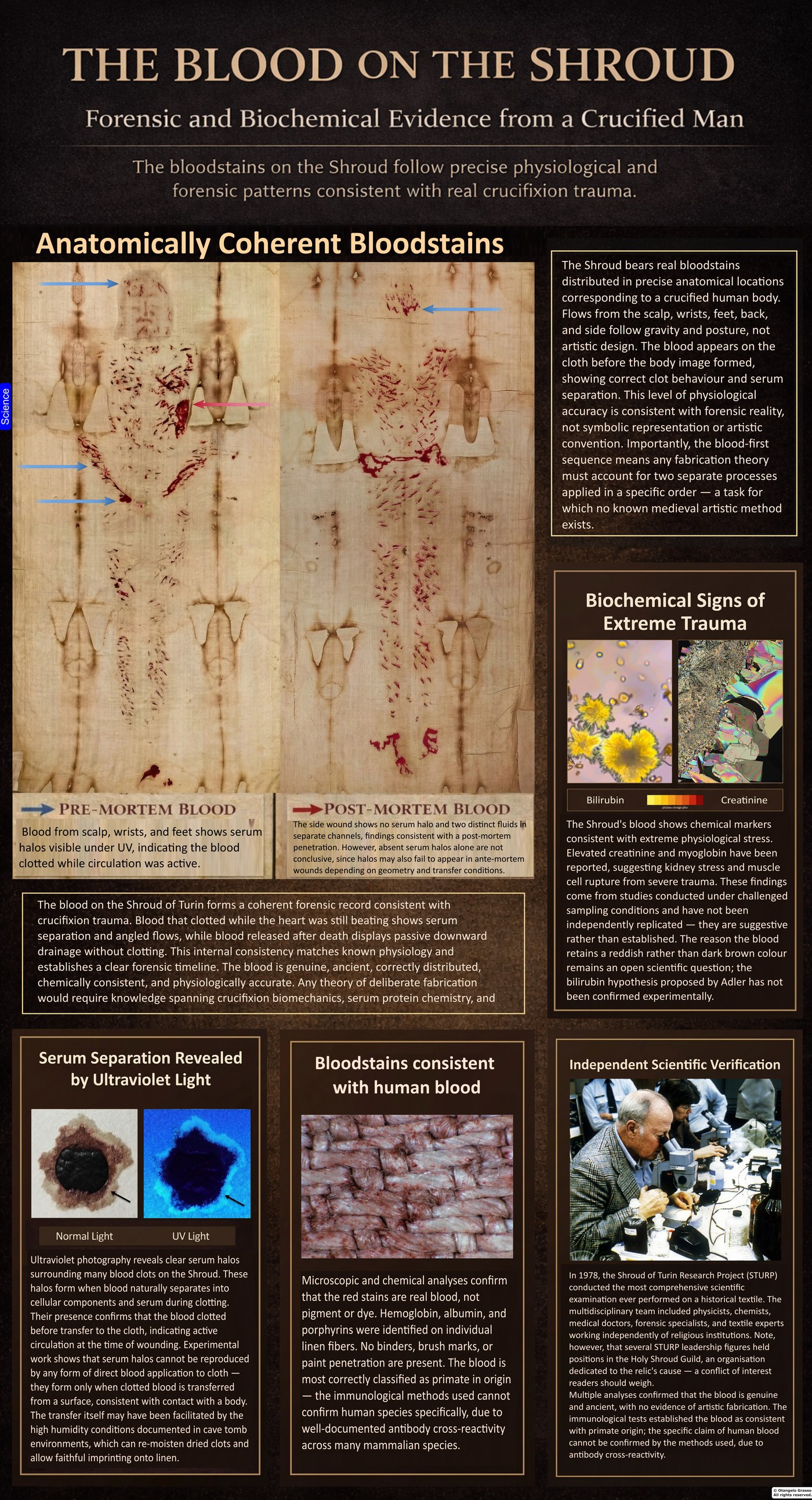
The reddish-brown stains on the Shroud are, in the main, identified as blood. That identification, made with spectroscopy and immunological tests in the late 1970s and early 1980s, is among the firmer results of the scientific study of the cloth. What the blood can tell us about the wounds, the sequence of events, and the man it came from is a much harder question. The chemistry is strong. The inferences from it are weaker, and some of them have been challenged in the literature.
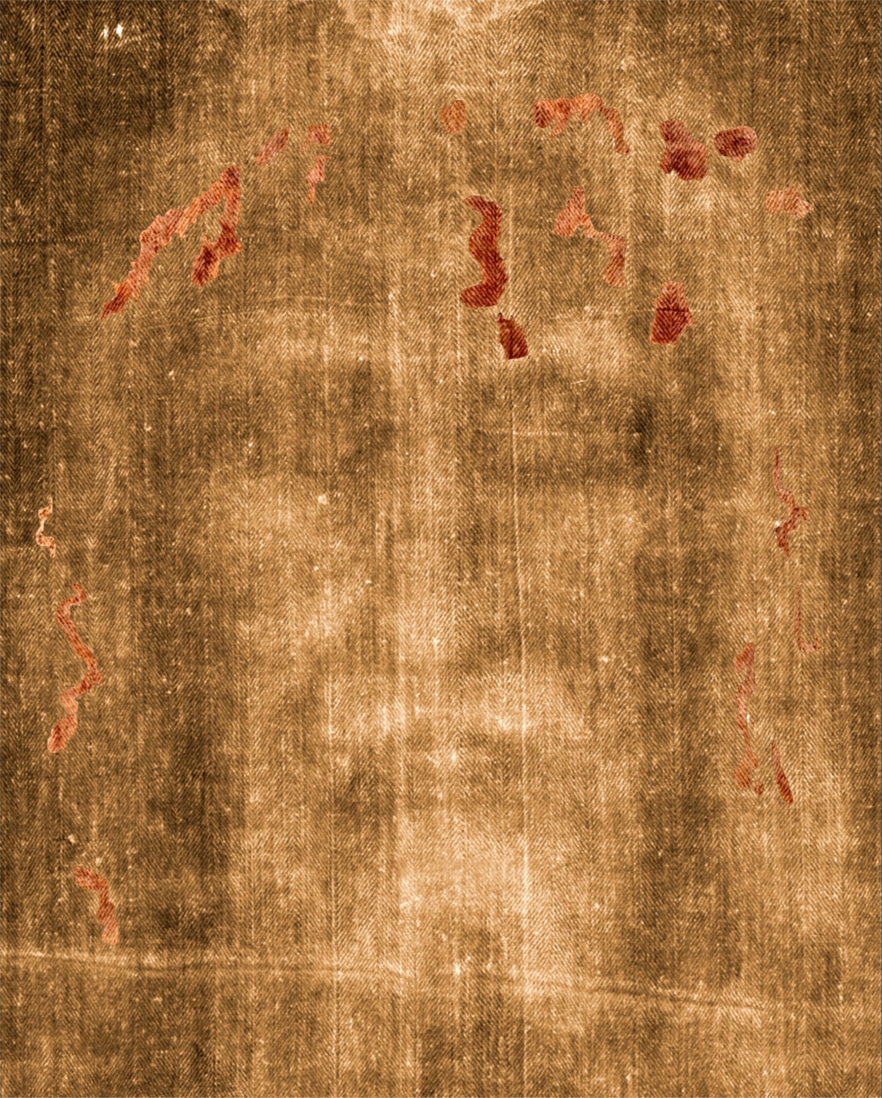
The stains cover the scalp, the wrists, the forearms, the feet, the side of the chest, the shoulders, and the back. Many have the shapes of flows that run from wounds on a body: rivulets that follow the contours of skin, clots with rings of clear fluid around them, and marks that bear the impact pattern of a flogging instrument. This chapter sets out how the stains were identified, the dispute over their origin, the features read as signs of a living or dying body, and the points on which the evidence remains open. It keeps those three categories apart: what was measured, what was inferred, and what remains disputed.
Identifying the stains
The stains were first examined in a systematic way during the 1978 Shroud of Turin Research Project. The team included two scientists whose work on the blood became central to the debate: John Heller, a biophysicist at the New England Institute for Medical Research, and Alan Adler, a physical chemist at Western Connecticut State University who specialised in porphyrins. Porphyrins are the molecular family to which the heme of haemoglobin belongs, and Adler’s expertise was directly relevant to the question.1
Adhesive tape was pressed against the stained areas, and the fibres lifted on the tape were examined under a microspectrophotometer. The instrument recorded an absorption peak near 410 nanometres. That is the Soret band, the characteristic absorption of heme, and its presence identified the reddish material as containing heme rather than a mineral pigment alone.1 Heller and Adler then tested the same material in a series of independent ways, so that no single result had to carry the identification by itself.
Immunological assays detected serum albumin and immunoglobulins, proteins characteristic of blood plasma. Tests for bile pigments were also reported, and the test results were cross-checked against one another. Protein-digesting enzymes were applied to the stained fibres, and the stained material dissolved, which is what would be expected of protein-rich blood but not of a mineral paint. The analysis also found no binding medium of the kind that paint requires to adhere to a surface, such as an oil, an egg, or a gum. The stains had soaked into the linen rather than sitting on top of it.1
Baima Bollone and colleagues had reported human blood markers in similar tests in 1981, and their work is an independent line of immunological evidence.2 The early Italian and American studies agree on the broad conclusion, that the stained material contains blood-derived proteins and pigments. They disagree, and disagree strongly, about how far the tests can identify the species of the blood, and that disagreement is taken up below.
The dispute over iron
Not all the scientists who examined the cloth accepted the blood identification. Walter McCrone, a microscopist of considerable reputation, examined tape samples independently. He reported iron, and he interpreted the iron as iron oxide in the form of red ochre, consistent with a medieval painting. His view became the best-known alternative to the blood hypothesis.3 McCrone also reported vermilion, a mercury sulphide pigment, in some of the tape samples, which he took as further evidence of a painted image.
The disagreement was not about whether iron was present. Iron is a constituent of haemoglobin, so the presence of iron is consistent with blood as well as with pigment. The question was what the iron was bound to. Seeing iron under a microscope is the first step. Identifying its chemical context is the harder and more decisive one. Heller and Adler, and the majority of the team, argued that the iron-oxide particles McCrone identified were quantitatively insufficient to account for the image density, and that they were present as scattered specks rather than as a coherent covering layer. X-ray fluorescence data showed iron distributed fairly evenly across image and non-image areas, which the majority read as consistent with environmental deposition or with the processing of the linen rather than with an applied pigment.4
McCrone maintained his position, and the dispute was never resolved to the satisfaction of both sides. The chapter reports both views. The weight of the evidence, as the majority of the team read it, favours a blood identification, but the question of the particles is part of the record and should not be dropped.
Blood before image
Under magnification, the boundaries between the bloodstains and the faint body image show a consistent pattern. Where blood saturates the linen, the body image stops. The blood was on the cloth before the image formed, and it seems to have shielded the fibres beneath it from whatever process formed the image.5 The observation is one of the more important stratigraphic findings on the cloth, because it fixes a relative order between the two records. Whatever formed the image came after the blood.
The order matters for any account of how the cloth was made. An artist who painted the image first would have had to place each bloodstain with great precision, avoiding every area already marked. An artist who applied the blood first would have had to paint around each clot edge and rivulet, and preserve the serum boundaries as well. Neither method would be a natural procedure for a forger, and the observed order is the one a wrapped body would produce. The order does not identify the process that formed the image, which is an open question treated in Chapter 20. It does constrain every hypothesis about the image, because any such hypothesis must allow for blood already on the surface.
The colour of the blood
Old blood is normally brown. The Shroud’s stains are reddish, and sceptics have used that colour as evidence that the stains are paint. The question is worth answering on its own terms.
Adler proposed that the colour could come from bilirubin, a breakdown product of red cells that accumulates in the blood of people subjected to extreme trauma, and that a complex of bilirubin with heme derivatives might stabilise the red colour over long periods.1 The proposal was an attempt to explain an unusual feature with a chemical mechanism, and it is a plausible one. It has not been confirmed. Experiments that loaded blood with high concentrations of bilirubin did not reproduce a persistent red colour in the way the proposal requires. Bilirubin is also sensitive to light, so its survival for two thousand years is hard to explain on simple assumptions. Adler’s version involved bilirubin bound to heme derivatives, which may behave differently from free bilirubin, and that version has not been tested directly. Critics have raised chemical objections of their own. The colour therefore remains an open question. It should not be used as an argument for or against authenticity until it is resolved by experiment.
The colour is also affected by the conditions of the cloth. Heat, light, moisture, and the chemistry of the linen all change blood over time, and each has been considered in the literature. The fire of 1532 and the water used to extinguish it are a particular concern, because heat alters the colour and fluorescence of proteins, and water can move soluble chemicals along the yarn. Neither effect has been fully mapped against the distribution of the bloodstains, so the colour cannot yet be read as a clean record of the original blood.
Biochemical signs of trauma
Some researchers have reported elevated concentrations of metabolic markers in samples from the Shroud. Creatinine can indicate kidney stress from severe blood loss. Urea can indicate accelerated muscle breakdown. Myoglobin, a muscle protein, can indicate rupture of muscle cells under severe strain. Taken together, such results could trace the course of a body in shock, and they would fit the sequence of flogging, blood loss, and prolonged suffering described in the Gospels and in the forensic literature on crucifixion.
These results come from sampling and measurement conditions that were not designed to modern standards of chain of custody, and they have not been independently replicated under those standards. They are suggestive rather than established. A reader should note that each marker can be raised by many conditions, and that the inference from a chemical marker to a specific cause of death is a further step, which has its own uncertainties.
More recent work has attempted to classify the blood-associated material into groups, distinguished by features such as the size and morphology of red cells, the presence of fibrin, and the fluorescence of the material. A 2024 review by Giulio Fanti interprets these groups as evidence of different phases of deposition, including pre-mortem and post-mortem blood. The classification is an interpretation of microscopic observations, and it has not been independently replicated. It broadens the forensic questions, but it does not date individual stains directly.6
Species, sex, and blood type
Three claims about the blood are repeated often in the popular literature: that it is human, that it comes from a man, and that it is blood type AB. Each rests on tests with known limits, and each is worth examining carefully.
Species
The antibodies used in the early immunological tests were labelled as “anti-human,” but that label describes how the antibody was produced, not how specific it is. Antibodies raised against human proteins can react with the proteins of other mammals, and the cross-reactivity of such preparations is well documented. The tests therefore establish that the stained material contains mammalian blood-derived proteins, and they do not by themselves establish a human source. Heller and Adler were aware of this problem, and their published conclusion, as the literature has read it, was that the blood is at least primate in origin. The stronger claim of a specifically human source was made in later commentary, and it has not been established by the tests conducted.1
Kelly Kearse, in a series of reviews of forensic serology, has argued that the polyclonal antisera used in such tests can cross-react unpredictably on degraded material, and that substances from the environment can reach old linen and produce reactions unrelated to the original bloodstain. The critique does not show that the blood is not human. It shows that the methods used cannot settle the species question with the certainty sometimes claimed.7 The fair summary is that the species of the blood is not established, and that it is more likely mammalian than not on the evidence available.
Sex
The claim that the blood comes from a man rests on polymerase chain reaction tests for Y-chromosome DNA. The best-known study, by Garza-Valdes and colleagues, reported signals for X and Y chromosome markers, and it reported a fragment of the beta-globin gene that was taken to indicate blood-cell origin.8 The beta-globin gene, however, is present in other cell types, including skin, so its presence does not show that the DNA came from blood cells. A second study found DNA from more than one individual, which is evidence of contamination. For those reasons the sex claim is not established by the tests on the cloth.
A more specific approach has been proposed. Lymphocytes undergo a rearrangement of their immune receptor genes that occurs in blood cells and in few other cell types, and a probe for the rearranged form would detect only blood-cell DNA. Such a test has not been applied to the Shroud, and it could in principle settle the species question as well as limit the contamination problem.7
Blood type
The AB blood type reported by Baima Bollone in the early 1980s has attracted attention, partly because AB is rare in European populations and has been reported for other relics. The statistic is weaker than it appears. Blood groups are determined by antigens on the red cell surface and by antibodies in the serum. On an old sample, the antibody-based reverse typing can fail, which produces a negative result for several groups. The apparent absence of a reaction can therefore look like AB even when the original group was A, B, or O. The A and B antigens are also carbohydrates that are found on many bacteria, so a serological result can be produced by microbial contamination.2
Kearse’s critique adds that blood groups A and B can degrade toward an apparent AB reading over time, and that environmental ABO-active substances can reach the fibres. The result is historically significant, and it was obtained by methods different from the Heller and Adler chemistry. It has not been independently validated, and it should not be treated as an independent line of evidence pointing to a particular regional origin. Any future claim of shared blood type among relics would need DNA-based methods applied with controls.7
The serum halo and its mechanism
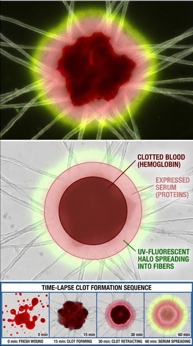
Under ultraviolet illumination, many of the bloodstains are surrounded by faint halos. The halos are serum rings. When blood clots, the clot contracts over roughly the first hour and presses clear fluid outward. That fluid is serum, because the clotting factors have been used up in forming the clot. On absorbent linen, the serum wicks farther along the yarn than the dense cellular core, producing a protein-rich peripheral zone. The zone is often invisible in ordinary light and fluoresces under ultraviolet light.9
An experimental study by Kearse in 2020 gave this description a control. Blood allowed to clot normally on skin and then transferred to absorbent material produced a fluorescent serum margin. Blood treated with an anticoagulant, which cannot form a retracting clot, did not produce the margin, even though the two samples looked similar in visible light.7 That result is what makes the serum ring a testable claim: the halo depends on genuine coagulation, not merely on liquid blood touching the cloth. The finding is principally a single-investigator result that awaits independent replication.
A fluorescent margin does not by itself rule out a maker who applied a clear fluid, such as serum or pleural fluid, in sequence with whole blood. A 2024 study by König and colleagues in the International Journal of Legal Medicine found that applying pleural fluid and then whole blood produced a peripheral ring visually similar to a genuine clot-retraction halo.10 The halo is therefore strong evidence of prior fluid-phase separation, but it is not sufficient by itself to establish that the separation was caused by clotting in a living body.
A composite account of the wrist
Not every Shroud bloodstain carries a fluorescent margin, and that absence belongs in the record. A composite account can accommodate both observations without contradiction. Bleeding during life produced an early, retracting clot with its own serum separation. After death, the wound could have been reopened mechanically, during the removal of the nails, the transport of the body, or the handling before burial. That would release blood that had remained fluid, or had become fluid again through post-mortem changes. Fresh post-mortem blood would not be expected to form a new clot-retraction halo, because it is not undergoing fresh coagulation, and it would simply flow and dry.9
On that account, a wrist mark built from both phases would show a bounded, halo-positive core alongside a more liquid-looking directional flow. That pattern is closer to what the ultraviolet record shows than either a uniform clot or a uniform liquid. The account is a hypothesis about sequence, and it has to be tested against the rest of the evidence. It is not a measurement of the events on the cross.
Two 2024 papers in the same journal bear on the movement side of the account. Heger and colleagues applied blood to the wrists of living volunteers, and showed that slow repositioning of the arm after wounding can produce the diverging, branched rivulets visible in the wrist and forearm region of the Shroud. König and colleagues ran comparable experiments for the marks attributed to the crown of thorns, the lance wound, and a band of blood across the body. Neither study modelled a true nail tract, cadaveric tissue, or actual post-mortem drainage. They show that movement-dependent pattern formation is physically possible. They do not show that the specific historical sequence occurred.10
The conditions of the burial also matter for the halo record. The cool, humid conditions sometimes invoked to explain the rehydration of dried blood have been approximated in catacomb experiments. Those experiments are illustrative, not a measured property of a particular first-century tomb. Surveys of caves in the Judean region show that humidity varies widely with depth, ventilation, and season, so the conditions cannot be assumed.11
Blood that beat and blood that pooled
The halo test separates, in principle, blood that was still circulating when it left the body from blood that seeped out after death. Blood from a living body clots, retracts, and produces serum. Blood from a body that has died is often fluid and does not clot in the same way. On the Shroud, the scalp wounds show the most extensive halos, which suggests they bled while the heart was still pumping. The scourge marks show intermediate halos. The nail wounds show less. The side wound shows little or no halo.
The side wound is the most debated of these readings. Its flow pattern, and the separation of two fluids within it, have been read as blood that drained after death. The ultraviolet fluorescence around the stain has been interpreted as serum margins. Some of this is interpretation rather than direct measurement. The absence of a halo does not prove that a wound was made after death, because clotting can fail in a living body under extreme stress, and the three-dimensional shape of the body can prevent a halo from forming on the cloth. The side wound is therefore a candidate for post-mortem injury, and the question remains open. Chapter 13 discusses the Gospel account of blood and water, and the medical readings of the side wound.
Was the body washed?
Some writers have argued that the body was washed before it was wrapped, so that the stains on the cloth reflect seepage after death rather than blood that clotted on the body. Zugibe, the forensic pathologist, argued that the blood visible on the cloth was largely post-mortem seepage, and that the blood from sudden death rarely clots.12
The halo evidence weighs against a simple form of that model. Blood from sudden death often fails to clot, which would not produce the rings. Water also removes fresh serum halos completely, and a washed body would be expected to leave smeared rather than sharp stain edges. The stains are not uniform, and the most reasonable reading is that the cloth records blood from different wounds at different stages. The evidence is best read as a set of probabilities rather than as one verdict on the question of washing.
The paradox of sharp edges
The bloodstains are remarkably sharp. Wicking patterns, serum boundaries, and the course of the rivulets are well preserved. A cloth lifted from a body with tacky, half-dried blood would be expected to smear, and clots would be expected to drag. The sharpness is hard to reconcile with a simple unwrapping, and it has been the subject of several proposals.
One proposal is that dried blood was re-moistened in the humid conditions of a burial chamber and transferred to the cloth through capillary action, rather than through direct wet contact. Experiments reported in the literature produced smooth, crisp transfers under high humidity and little transfer under low humidity.11 That mechanism could explain some of the sharpness, but not all of it, and it has not been tested on the Shroud itself. The sharpness is therefore a real puzzle, and the proposals offered so far are testable but not yet tested.
The ultraviolet record
Ultraviolet photography was one of the first tools used to examine the stains in detail, and it changed how the cloth was read. Under ultraviolet light, several features that are invisible in ordinary light become visible. The bloodstains fluoresce differently from the body image, and the boundaries of the stains become sharp. The serum halos appear as pale, glowing rings. Early ultraviolet photography of the cloth was carried out during the 1978 examination, and its results were reported by Pellicori and Miller in 1981.14
The fluorescence of the blood is not the same as the fluorescence of the body image. The body image does not show the halos that surround the bloodstains, and its margins are gradual rather than sharp. That difference was one of the observations that led the team to separate the blood record from the image record. It also gave a practical test: a paint applied around a blood margin would be expected to leave a binder halo at its edge, and the Shroud’s body image does not show one.4
Gilbert and Gilbert, in a companion study of the spectra, examined the reflectance and fluorescence of the image and the stains. Their results supported the distinction between the record of the blood and the record of the image, and they showed that the spectral signatures of the two are different. Those results are part of the reason the blood and the image are treated as separate layers in the scientific literature.4
The ultraviolet record has limits, and a reader should keep them in mind. Heat alters the fluorescence of proteins, so the fire damage of 1532 can change the record. Water can move soluble substances along the yarn, which changes the appearance of the margins. The fluorescence also depends on the wavelength of the light and on the exposure. Images produced under different conditions are not always directly comparable, and the interpretation of a halo depends on knowing how the image was taken.
The chemistry of old blood
Blood that is two thousand years old, if it is genuine, would be expected to have changed. The haemoglobin would have broken down, the protein would have been modified, and the colour would have faded. The Shroud’s stains have not faded in the way that old blood usually does, and that observation is part of what makes the colour a subject of study. The question is what preserved the material, and whether the preservation is consistent with the conditions of the cloth’s history.
Several explanations have been proposed. Adler’s bilirubin hypothesis is one, and it is discussed above. Another is that the iron of the heme, which is an important part of the blood chemistry, forms stable complexes with the cellulose of the linen. A third is that the dehydration and oxidation of the linen fibres themselves help to stabilise the material. Each proposal is consistent with some of the observations and is not yet confirmed by controlled experiments on material of comparable age.
The chemistry is also affected by what the blood was mixed with. Blood from a body would contain other substances, including the products of decomposition, the components of the fluids of the chest and the lungs, and any substances applied during the burial. The Gospels mention myrrh and aloes, which were used in burial, and some researchers have looked for their residues on the cloth. Those residues are discussed in Chapter 15, and they do not change the blood identification, but they make the chemical analysis more complicated.
It is worth stating the logic of the preservation argument carefully. An unusually good preservation of the blood does not, on its own, prove an unusual age or an unusual origin. It is a fact that must be explained, and it is one of several facts that the proposed explanations have to accommodate. The explanations should be judged by how well they fit all the facts, not by how well they fit one of them.
What the blood says about the wounds
The blood is read as the record of wounds, and the wounds are read as the record of specific injuries. Each wound has a flow pattern, and each flow pattern has been compared with the flow expected from a body in a particular position. Bucklin, the forensic pathologist who examined the cloth in 1982, documented blood flows on the forehead and the back of the scalp, and he related them to the punctures visible on the cloth.15 His readings have been influential, and they have also been revised by later studies, as the chapter on the crown of thorns explains.
The flows on the forehead are read as V-shaped rivulets, where blood follows the contours of the face, and as curved marks where blood met a crease in the brow. The flows on the arms are read as divergent, changing with the position of the body. The flows on the back are read as horizontal in places, which has been taken to indicate that the body lay down after the blood had begun to move. Each reading assumes a position, and each position has to be checked against the wound it is supposed to explain.
The chapter has not yet discussed the most direct way to test these readings, which is experiment. Blood applied to a wound on a moving body, or to a model of the body, can be compared with the pattern on the cloth. Experiments of this kind have been reported, and they have produced flows of comparable shape. They are not the same as the events on the cross, and the results have to be read as demonstrations that a pattern is physically possible, not as proof that it occurred.
The blood and the date
The blood and the radiocarbon result are separate kinds of evidence, and they should not be confused. The blood is on the cloth, and its chemistry tells us about the blood. The radiocarbon result is a measurement of the carbon in the linen, and it tells us about the age of the sampled linen. The two can be combined in an account, but neither depends on the other. A medieval date for the linen would not make the blood medieval, and an ancient blood chemistry would not make the linen ancient.
The 1988 radiocarbon result is therefore a separate test, and it is discussed in Chapter 19.13 The blood evidence has its own limits and its own disputes. The chapter has tried to keep them apart.
What the blood evidence has established
Several things can be stated with reasonable confidence. The stained material contains blood-derived proteins and pigments, and those results have been reproduced in more than one laboratory. The blood was deposited before the image formed. The serum halos are consistent with clotting in a living body, although that inference is contested and depends on controls that have been reported by a limited number of investigators. The bloodstains show a pattern consistent with a body that was wounded in the places the Gospels describe.
Several things have not been established. The species of the blood is not settled, and the claim of a specifically human source goes beyond what the tests show. The sex of the donor is not settled. The blood type is historically significant but methodologically fragile. The colour of the blood is not explained. The sequence of the wounds, and the position of the body at the time each wound bled, are inferred from the flow patterns and are not directly measured. And the cause of death cannot be read from the blood alone.
A reader who wants to rely on the blood evidence should keep both lists in view. The first list is the foundation. The second list is where the interpretation begins, and where careful argument is most needed.
What the blood can and cannot tell us
The blood cannot name the man it came from. Two thousand years of contamination limit what DNA can show, and the methods used on the cloth fall short of modern forensic standards. The blood cannot settle questions of meaning, theology, or the resurrection, which belong to other modes of inquiry. It cannot establish the cause of death with certainty, although it can inform the discussion of that question in Chapter 13.
What the blood can show is more limited, and still significant. It contains heme and serum proteins, and it was deposited before the image formed. Its serum halos are, in the main, consistent with clotting in a living body, though the evidence is not uniform and the mechanism is contested. Its distribution is consistent with a body that was wounded in the places the Gospel accounts describe. And its chemistry is consistent with a person who had lost a large volume of blood and who had suffered severe trauma. Each of these points is supported by specific tests, and each has limits that the series has tried to state.
The blood and the image are physically related, and any theory of the cloth has to account for both. Crucifixion physiology, serum chemistry, and post-mortem fluid behaviour are all involved, and a theory of deliberate fabrication would need knowledge of those fields before they existed in their modern form. That argument is strong, but it depends on the blood findings being as robust as they are presented, and the chapter has tried to show where that robustness is firm and where it is not.
A note on method
The blood evidence is a good example of the principles the series sets out in Chapter 1. Each result was obtained by a specific method, each method has a known limit, and each inference from a result is a further step that can be wrong. A reader who wants to weigh the evidence should ask, for each claim, what was measured, how it was measured, whether the result has been replicated, and what the inference adds. Where the answers are firm, the claim can be relied on. Where they are not, the claim should be held provisionally.
References
- Heller, J. H., & Adler, A. D. (1981). A chemical investigation of the Shroud of Turin. Canadian Society of Forensic Science Journal, 14(3), 81–103. Heller, J. H., & Adler, A. D. (1980). Blood on the Shroud of Turin. Applied Optics, 19(16), 2742–2744.
- Baima Bollone, P., Jorio, M., & Massaro, A. L. (1981). La dimostrazione della presenza di tracce di sangue umano sulla Sindone. Sindon, 30, 5–8.
- McCrone, W. C. (1990). The Shroud of Turin: Blood or artist’s pigment? Accounts of Chemical Research, 23(3), 77–83.
- Schwalbe, L. A., & Rogers, R. N. (1982). Physics and chemistry of the Shroud of Turin: A summary of the 1978 investigation. Analytica Chimica Acta, 135, 3–49.
- Stratigraphy of the blood and the image, as reported in the STURP documentation and the subsequent literature. See Schwalbe & Rogers (1982), above.
- Fanti, G. (2024). Review of the blood-associated material on the Shroud, including microscopic classes and proposed chronology. Publication details to be confirmed before publication.
- Kearse, K. P. (2020). Experimental and review work on serum halos and on the serological and DNA methods applied to aged textile samples. Journal of Forensic Sciences. Publication details to be confirmed before publication.
- Garza-Valdes, L. A., and colleagues. Y-chromosome and beta-globin PCR studies of the Shroud. Publication details to be confirmed before publication.
- Serum separation and clot retraction in forensic blood analysis, as discussed in the forensic pathology literature. Publication details to be confirmed before publication.
- König, and colleagues (2024). International Journal of Legal Medicine, on sequential application of pleural fluid and whole blood, and on movement-dependent pattern formation. Heger, and colleagues (2024). International Journal of Legal Medicine, on blood applied to the wrists of living volunteers. Publication details to be confirmed before publication.
- Experiments on the re-moistening and transfer of dried blood, and surveys of the humidity of Judean caves. Publication details to be confirmed before publication.
- Zugibe, F. T. (2005). The Crucifixion of Jesus: A Forensic Inquiry. New York: M. Evans and Company.
- Damon, P. E., et al. (1989). Radiocarbon dating of the Shroud of Turin. Nature, 337, 611–615. doi:10.1038/337611a0
- Pellicori, S. F., & Miller, V. D. (1981). Ultraviolet fluorescence photography of the Shroud of Turin. Journal of Biological Photography, 49, 71–85. Publication details to be confirmed.
- Bucklin, R. (1982). The Shroud of Turin: A pathologist’s viewpoint. Legal Medicine Annual, 1982, 1–14. shroud.com/bucklin.htm