The Shroud of Turin · Chapter 18 of 21
The interface: the STURP investigation and the limits of forensic science
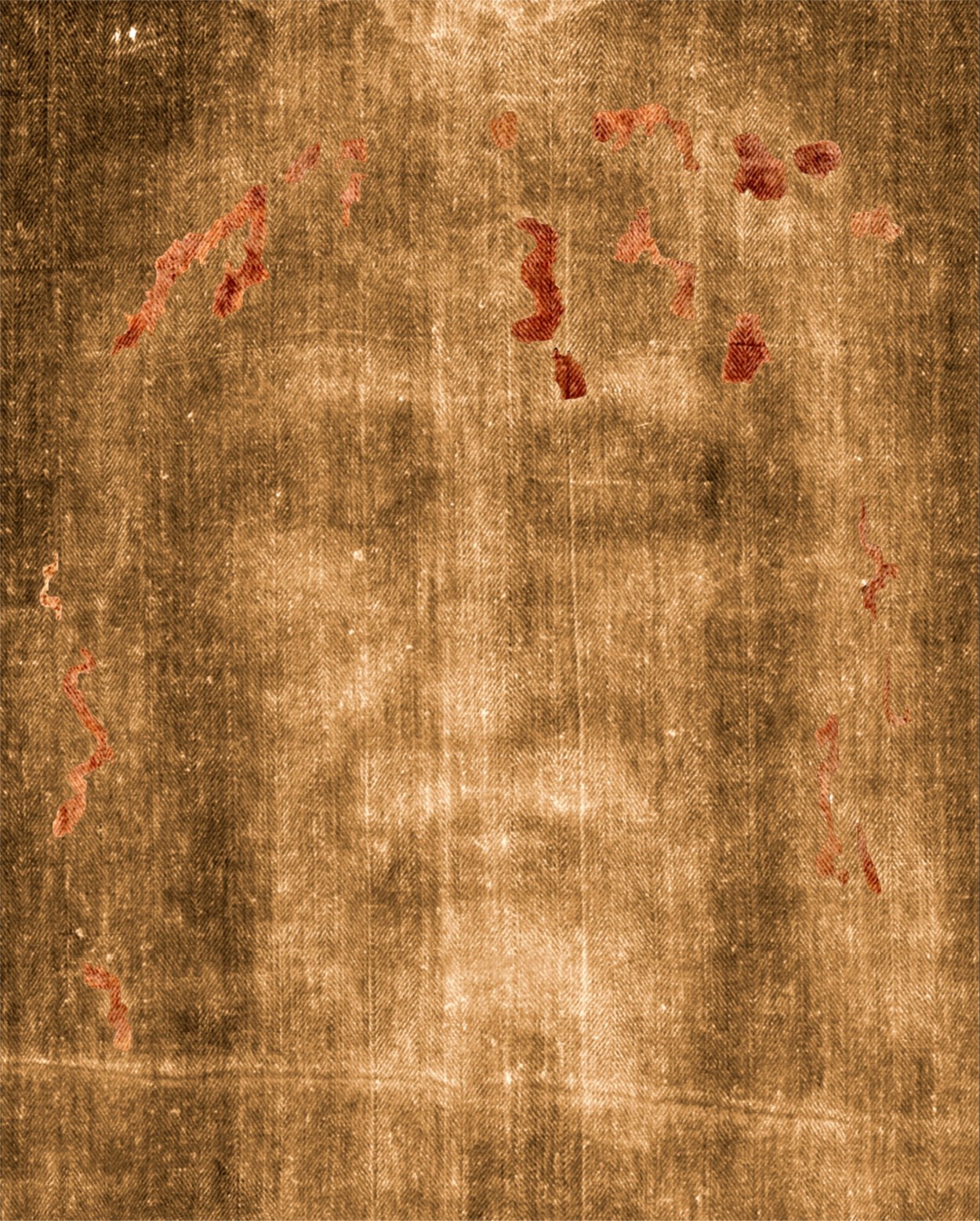
In October 1978 a team of about thirty-three scientists, engineers and technicians spent five days and nights examining the Shroud of Turin in the cathedral of Turin. They worked under strict conditions. The cloth was in a sealed case, the examination was carried out at night, and the team had only a few days to make its observations. They brought image analysers, ultraviolet cameras, microscopes, spectrometers and a set of sticky tapes for sampling the surface. Their aim was to answer questions that had been argued for decades: what the image was, what had made it, and whether the stains on the cloth were blood. The team was called the Shroud of Turin Research Project, and it is known by the acronym STURP. This chapter sets out how the team worked, what it found, what it did not claim, and how its results were received. It also sets out the criticism that followed, and the limits of what forensic science can establish about an object of this kind.
Why the 1978 project was formed
The Shroud had been examined before 1978, most notably by Secondo Pia in 1898 and by Giovanni Enrie in 1931, both of whom made photographs of the cloth. The photographs showed the image as a negative, and they gave the first clear view of the face. The 1931 photographs were more detailed, and they were widely studied. Later examinations, including the work of Pierre Barbet in the 1930s and the Turin examinations of the 1970s, added more information. By the late 1970s the image had been photographed many times, but no team had yet brought the full range of modern instruments to bear on the cloth.
The project was initiated by a group of American scientists who were interested in the image and who had access to modern equipment. The group included John Jackson, a physicist and a leader of the project, and Eric Jumper, an engineer who worked with the image analyser. The project was organised as a team of specialists, with different groups responsible for different tests. The team’s members came from academic, industrial and government laboratories, and they worked on the project as volunteers, with funding from private sources.
The conditions of the examination
The examination was carried out under conditions set by the Church and the custodians of the cloth. The team was allowed to work for a limited period, and the cloth was not to be removed from its case or subjected to any treatment that would damage it. The examination was carried out at night, when the cathedral was closed to visitors, and the team worked in shifts. The cloth was kept at a controlled temperature and humidity, and the team took steps to avoid contamination.
The conditions shaped the results. The team could not take the cloth out of its case, and could not sample it in the ways that a laboratory would have liked. The examination was therefore a survey, not a full investigation, and the team was clear about the limits of what it could do. Those limits are important for reading the results, because many of the questions that remained open after 1978 were questions that the conditions had not allowed the team to answer.
The instruments
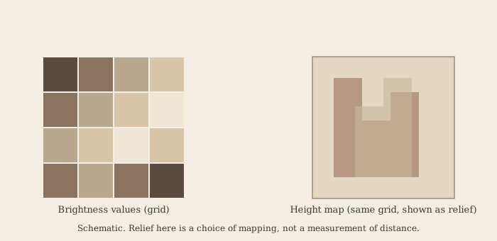
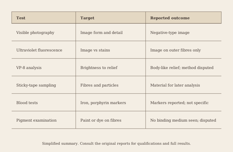
The team used a wide range of instruments. They included visible-light photography at high resolution, ultraviolet photography and fluorescence, infrared photography, X-ray imaging, microscopy, spectrometry and the VP-8 image analyser. The VP-8 was designed to convert the brightness of a photograph into a height map, and it had been developed for the analysis of aerial and satellite images. The instruments were chosen to examine the cloth at different scales and with different physical principles, so that the results could be compared.
The use of many instruments was a strength of the project, because it allowed the team to check one result against another. It also created a problem, because the instruments measured different things, and their results could not always be combined into a single account. The team had to decide how to weigh each result, and the interpretation of the combined data was a matter for judgement as much as for measurement.
The VP-8 image analyser
The VP-8 was the most discussed instrument in the project. It was built to process images of the earth and of the surface of other planets, and it could take a photograph and produce a map of the brightness of each point. The brightness could then be converted to a height, using a fixed transfer function. The result was a three-dimensional representation of the image, which showed the face and body as a relief.
The VP-8 analysis of the Shroud was first carried out in 1976, by Jackson and Mottern at Sandia Laboratories, using a photograph of the cloth. The result suggested that the image contained information about distance, in the sense that brighter areas of the image were closer to the cloth, and darker areas further away. The team in 1978 repeated the analysis, and they reported that the relief was consistent with a body. The analysis was an important point in the argument for the image’s uniqueness, and the chapter discusses it further below.1
The ultraviolet work
The ultraviolet examination was among the most important of the 1978 observations. The team shone ultraviolet light on the cloth and photographed the fluorescence of the surface. The image on the cloth appeared dim under ultraviolet, and the areas that were blood-stained showed a different fluorescence from the linen. The stains absorbed the ultraviolet light, and the difference in response was used to distinguish the stains from the image.
The team reported that the image itself showed a fluorescence that was limited to the outermost fibres of the linen, and that the image did not penetrate the thread. The observation has been central to the argument that the image was formed by a process that affected only the surface of the fibres, and not by a pigment that soaked into them. The observation also bore on the question of whether the image was a painting, because a painting would be expected to show a different fluorescence pattern.
The sticky-tape samples
The team collected samples from the cloth using sticky tapes. The tapes were pressed onto the surface of the cloth in selected areas, and then removed, carrying with them the loose fibres and particles from the surface. The samples were taken from the image, from the blood marks and from the background linen, so that they could be compared. The tapes were distributed to laboratories for analysis, and the results were reported in the following years.
The sampling method was carefully chosen, because the cloth could not be cut or damaged. The tapes gave a small amount of material, but the material was enough for many kinds of analysis. The method also had limits. The tapes could pick up particles from the air or from the surfaces they touched, and the samples could be contaminated by handling. The team was aware of these risks, and it used control samples to check for contamination.
The blood tests
The blood stains were tested by several methods. The team looked for the chemical markers of blood, including the iron of haemoglobin and the porphyrin that is a breakdown product of it. The tests were carried out on the tape samples and, in a few cases, on material taken with other methods. Some tests gave positive results for the markers, and others were negative or inconclusive.
John Heller and Alan Adler carried out a separate chemical investigation, which was published in 1981. They reported that the stains contained substances consistent with blood, including porphyrin, and that the stains were not consistent with paint. The results have been widely cited, and they have been disputed. The chemical tests are not specific to blood in every case, and the identification of the markers depends on the interpretation of the test results.2
The image without pigment
One of the central findings of the team was that the image did not contain a pigment or a dye of a kind that would be expected in a painting. The team examined the fibres under the microscope and reported that the colour of the image was carried by the outer surfaces of the fibres, and that there was no evidence of binding media or of paint applied in layers. The image was described as a fine discoloration of the linen, with a density that varied in a way that the team could not reproduce by painting.
The conclusion has been contested. Walter McCrone, a microscopist, argued in 1980 and later that the image contained iron oxide pigment in a form that suggested paint, and he reported particles of the pigment in the samples. The dispute between McCrone and the STURP team has continued, and it is one of the central disagreements of the Shroud literature. The reader should note that the question is whether the particles are the image, or whether they are contamination or background.3
What STURP concluded
The team did not issue a single conclusion that all members signed. The final statement was a summary of observations, and it described the image, the blood and the cloth in terms that were careful about their limits. The team said that the image was not a painting in the usual sense, that the blood stains were consistent with blood, and that the image had properties that the team could not reproduce by any method then available. It did not say who made the image, when, or how.
The team’s conclusions were therefore a set of observations, and not a verdict on the origin of the cloth. The team members held different views about the origin, and some of them remained open to the possibility that the cloth was medieval. The collective statement reflected that range of views, and it is the basis for the careful reading of the team’s results that the series adopts.
The radiocarbon sample and the 1978 team
The 1978 team did not take a sample for radiocarbon dating. The radiocarbon test was carried out in 1988, and it is discussed in Chapter 19. The team did, however, discuss the question of the sample. The team members argued that the cloth should be dated with a method that did not destroy a large part of it, and some of them argued that the dating should be carried out by several laboratories on the same sample. The argument shaped the protocol of 1988, which is also discussed in Chapter 19.
Criticism of the methods
The STURP methods were criticised on several grounds. The first criticism is that the examination was short, and that the team could not repeat the tests. The second is that the team members had a stake in the outcome, because some of them had previously expressed views on the authenticity of the cloth. The third is that the VP-8 analysis depended on the transfer function and the photograph, and that the relief was an artefact of the choices made. The fourth is that the sticky tapes were not controlled for contamination in every case, and that the blood tests were not specific enough to identify blood with certainty.
The criticism is not all of one kind. Some critics argued that the team was too sympathetic to the authenticity of the cloth, while others argued that the team was too cautious about the blood. The critics also differed about the pigment question and about the VP-8. The balance of the criticism shows that the team’s results were important, and that they were not beyond dispute.
The later replications
The STURP observations were tested in later years, both by members of the team and by other researchers. Some of the observations were confirmed, including the fluorescence of the image and the presence of the blood markers. Others were disputed or not replicated, including the relief of the VP-8 and the pigment analysis. The replications show that the observations are sensitive to the method and to the interpretation, and that the team’s results should be read with the limits in view.
Among the later scientific work, the 1984 paper by Jackson, Jumper, Ercoline and others, published in Applied Optics, gave a fuller account of the image analysis and the three-dimensional encoding. The paper set out the methods in more detail, and it stated the limits of the relief analysis. It is one of the more careful later publications, and it is cited throughout the series.4
What forensic science can establish
The STURP investigation shows what forensic science can establish about an object of this kind. It can describe the image, the stains and the cloth in detail. It can test some hypotheses about the image formation and the blood. It can show which hypotheses are inconsistent with the observations. It cannot, by itself, establish the date of the cloth, the identity of the person, or the process by which the image was made, except where a test is directly decisive.
The limits are important, because the Shroud has been the subject of many claims that go beyond what the observations can bear. A claim that the image was made by a particular process, or that the cloth was made at a particular time, must be tested against the observations. The series treats the STURP results as the best available record, and it treats the claims built on them with appropriate caution.
The interface between science and faith
The STURP team worked in a setting that was both scientific and religious. The cathedral and the custodians of the cloth were part of the setting, and the team members were aware of the devotional significance of the object. The team’s approach was to separate the scientific observations from the religious question, and to report the observations without claiming to answer the question of faith. The series adopts the same separation.
The interface between science and faith is not always easy to maintain. Some members of the public read the STURP results as a confirmation of faith, and some read them as a refutation. Neither reading follows from the observations alone, and the series asks its readers to keep the two questions apart.
Spectroscopy and the infrared work
The team used spectroscopy to examine the chemistry of the image and the stains without touching the cloth. Infrared reflectance spectroscopy measured how the surface reflected light at wavelengths beyond the visible range, and it could distinguish some materials from others by their characteristic absorption. Ultraviolet-visible spectrophotometry measured the absorption of light at shorter wavelengths, and it gave information about the colour-bearing compounds. The instruments were used on the cloth directly, and on the tape samples, and they gave the team a set of spectra to compare.
The spectra were not decisive on their own. Many organic materials have similar spectra, and the identification of a compound requires reference data and careful comparison. The team compared the spectra of the image with reference spectra of pigments, dyes, plant materials and blood-related compounds. The comparison excluded some common pigments, and it was consistent with the image being a discoloration of the linen. The team did not claim to identify the image compound, and it reported the spectral results as observations within a range of possibilities.
The infrared work also showed how the image behaved under different light. The image was visible in the infrared photographs, and its contrast differed from that in the visible photographs. That difference was used to argue that the image was not formed by a surface pigment, because a pigment would be expected to show a different infrared response. The argument has been accepted by some and questioned by others, and it depends on assumptions about the reflectance of the linen and the image.
The X-ray and the cloth structure
X-ray imaging was used to examine the structure of the cloth, and in particular the weave and the thread. The X-ray images showed the threads and the spaces between them, and they could reveal whether the cloth had been repaired or altered. The team used X-ray images to check the cloth’s structure against the photographs, and to look for signs of later additions.
The X-ray work also gave information about the materials. Some elements show more strongly in X-ray images than others, and the team looked for the presence of metals and other heavy elements that might indicate a pigment. The results were not consistent with a pigment-based image, and they suggested that the image was not made with a heavy-metal paint. The conclusion was qualified, because the X-ray detection depends on the amount of material, and small amounts may not be visible.
Microscopy and the fibres
The microscopic examination of the fibres was among the most detailed of the 1978 observations. The team looked at the threads under high magnification, and they described the surface of the fibres, the presence of any material on them, and the distribution of the image colour. The image colour was found on the outer surfaces of the fibres, and not in the interior, and the colour was described as a yellowing or browning of the fibre walls.
The microscopy showed that the image colour was not uniform along a fibre. The colour was denser in some places and thinner in others, and the variation was consistent with a process that acted on the surface. The team reported that the colour did not look like a layer of paint, and that the fibres showed no sign of the binding medium that would be expected in a paint. The observation was an important point in the argument against painting, and it has been contested by those who argue that the paint is present in small quantities.
Controls and contamination
The team was aware that its results could be affected by contamination and by the control samples. The control samples were taken from the cloth at places away from the image, and from other linen of similar age and type, so that the results could be compared with a baseline. The controls were used to check the tapes, the instruments and the handling, and they were part of the design of the study.
The controls had limits. The linen used as a control was not identical to the Shroud, and the background of the cloth was not uniform. The contamination of the tapes could come from the air, from the handlers, and from the surface of the cloth itself. A control could show the presence of a contaminant without showing its source. The team reported the controls and their limits, and the reader should weigh them when judging the results.
Dissent within the team
The team did not agree on every point. Some members were persuaded that the image could not be a painting, and that it was not made by any known process. Others were more cautious, and they argued that the observations were consistent with a medieval origin, provided that the process of image formation was not yet understood. The disagreement is important, because it shows that the collective statement was a compromise, and that the individual members held different views about the implications.
The dissent also concerned the blood. Some members were confident that the stains were blood, and others were less sure, because the chemical tests were not specific enough to identify blood with certainty. The team’s collective statement reflected a middle position, in which the stains were described as consistent with blood, and not as proven to be blood. The distinction is important for later arguments, and it should be kept in view.
The image without brush marks
The Shroud image shows no brush marks, no outlines of the kind that a painter would draw, and no evidence of corrections or changes. The image is continuous across the cloth, and its edges are soft. The team regarded these features as important, because they are difficult to produce by painting, and because a painter would normally leave some trace of the working process. The absence of such traces is one of the observations that has been used to argue that the image was not painted.
The argument has limits. A skilled painter working in a fine medium could leave few traces, and the absence of brush marks does not prove that no paint was used. The observation is an argument from absence, and its force depends on how difficult the painting would have been. The team did not claim that painting was impossible, only that the image did not show the characteristics of a painting as they would be expected to appear.
Replication attempts
The team and later researchers attempted to replicate the image. Some tried to produce a similar discoloration of linen by heat, by chemical treatment, or by radiation, and some attempted to produce the relief by painting. The attempts produced images that resembled the Shroud in some respects, but none produced the full set of features at once. The replications are useful because they show which features are hard to reproduce, and they set a standard for future claims about the image.
The replications also show the limits of replication. A failed attempt to reproduce the image does not prove that the image was made by a process that the attempts did not use. The attempts are tests of particular hypotheses, and each hypothesis must be stated before the test is carried out. The series treats the replication work as useful but limited.
The public reception
The STURP results were widely reported, and they reached a public audience that had little understanding of the methods. The reports often stated conclusions more strongly than the team had done, and they sometimes omitted the qualifications. The public reception shaped the debate, and it gave some claims an authority that the observations did not always support. The reader should be aware that the popular version of the STURP results is not the same as the team’s own statement.
The public reception also brought criticism from the scientific community. Some scientists questioned the methods, and others questioned the interpretation. The criticism was sometimes fair and sometimes unfair, and the reader should judge each claim on its merits. The series tries to report the criticism accurately, and to separate it from the claims it addresses.
What the STURP results do not decide
The STURP results do not decide the question of the Shroud’s date, its maker or its history. They describe the image, the stains and the cloth, and they set limits on the hypotheses that can explain them. They do not identify the process by which the image was made, and they do not say whether the image is the image of a crucified man. Those questions require other evidence and other methods, and the series treats them in the chapters that follow.
The practice run
Before going to Turin, the team carried out a practice run in September 1978, at a laboratory in Amston, Connecticut. The run allowed the team to test its procedures, its instruments and its communication. The members set up the equipment, ran the tests on reference materials and on test images, and timed each step against the limited hours available in Turin. The practice run also revealed weaknesses in the plans, some of which were corrected before the team arrived.
The practice run is a useful lesson in forensic method. A study with a short and fixed window must rehearse its procedures, because mistakes made under time pressure are hard to correct. The run also shows that the team was aware of the constraints, and that it tried to design its work so that the most important observations could be made in the time available. The reader who judges the STURP results should keep the constraints in mind, because they shaped the choice of tests.
The later debate over the pigment
The pigment question has been the subject of a long and bitter debate. McCrone and his colleagues argued that particles of iron oxide, in a form used in medieval paints, were present on the image, and that they could account for its colour. The STURP team and its supporters argued that the particles were background contamination, that they did not account for the image, and that the image was not a paint layer. The two sides used different sampling methods, different instruments and different criteria for identifying the particles.
The debate is useful for the reader because it shows how a single observation can be read in two ways. The particles are real, and their presence is not disputed. Their meaning is disputed, and the disagreement turns on whether they are the image or something else. The reader should look at the sampling, the controls and the criteria used by each side, and should ask which interpretation is better supported.
The critics of the methods
The critics of STURP included scientists who had not taken part in the project and who questioned its methods in print. Their objections focused on the sampling, the controls, the interpretation of the VP-8 relief and the identification of the blood markers. Some of the criticism was technical and well-founded, and it led to improvements in later work. Other criticism was broader, and it questioned the motives of the team members. The series treats the technical criticism as the more useful, and it notes the broader criticism where it bears on the results.
Closing the account of 1978
The STURP investigation remains the most extensive scientific examination of the Shroud. It produced a large body of observations, a set of careful limits on what those observations can show, and a standard of method that later work has tried to meet. It did not settle the questions of origin, date or authorship, and it was not intended to. Its lasting value lies in the precision of its descriptions and in the honesty of its statements about what remained uncertain.
What STURP contributes to the series
The STURP investigation is the foundation of the modern scientific study of the Shroud. Its observations are the starting point for most of the later work, including the chemistry, the image analysis and the dating. The next chapter takes up the radiocarbon test, which is the most publicised scientific result on the cloth, and which has been the subject of the most sustained dispute.
How to read the 1978 results
A reader who wants to use the STURP results should separate three kinds of statement. The first is the observation: what the team saw, measured or recorded on the cloth, such as the fluorescence of the image or the presence of material on the tapes. The second is the interpretation: what the observation is taken to mean, such as that the image is not a paint layer. The third is the conclusion: what follows about the origin, date or maker of the cloth. The observations are the firmest, the interpretations are contestable, and the conclusions go beyond the evidence unless further work supports them. Most of the public disagreement about STURP arises from treating an interpretation as an observation, or a conclusion as an interpretation. Keeping the three apart is the simplest discipline the series can offer, and it is applied throughout the chapters that follow. A reader who applies it to any claim about the Shroud, including the claims made in these pages, will know which parts of the argument rest on the cloth and which rest on the reader’s own judgement.
The reader should also note the date of each observation. The 1978 examinations were made with the instruments of that period, and the later work has improved some of them. Where a later study has repeated a 1978 observation with better equipment, the later result should be preferred, and where the later result differs, the difference is itself information.
The STURP investigation therefore sits at the boundary between what can be measured and what must be inferred. The team measured the image, the stains and the structure of the cloth with instruments that were advanced for their time, and it reported those measurements with care. Where it moved from measurement to inference, its members disagreed, and the collective statement stopped short of the conclusions that some of them held. That restraint is part of the value of the work. A reader who takes the STURP results seriously will accept the observations as the firmest ground the series has, while keeping the interpretations open to the same scrutiny as any other claim. The next chapter turns to the one scientific test that was designed to answer the question of date directly, and whose result has been the most publicised and the most disputed of all.
References
- Jackson, J. P., Jumper, E. J., & Ercoline, W. R. (1984). Correlation of image intensity on the Turin Shroud with the 3-D structure of a human body shape. Applied Optics, 23(14), 2244–2270. doi:10.1364/AO.23.002244
- Heller, J. H., & Adler, A. D. (1981). A chemical investigation of the Shroud of Turin. Canadian Society of Forensic Science Journal, 14(3), 81–103.
- McCrone, W. C. (1999). Judgment Day for the Shroud of Turin. Amherst, NY: Prometheus Books. The author’s pigment analysis and his critique of the STURP results. Edition and page details to be confirmed before publication.
- Jackson, J. P., Jumper, E. J., Ercoline, W. R., et al. (1984). The three-dimensional image of the Shroud of Turin. Applied Optics, 23(14). Page range and author list to be confirmed against the original before publication.